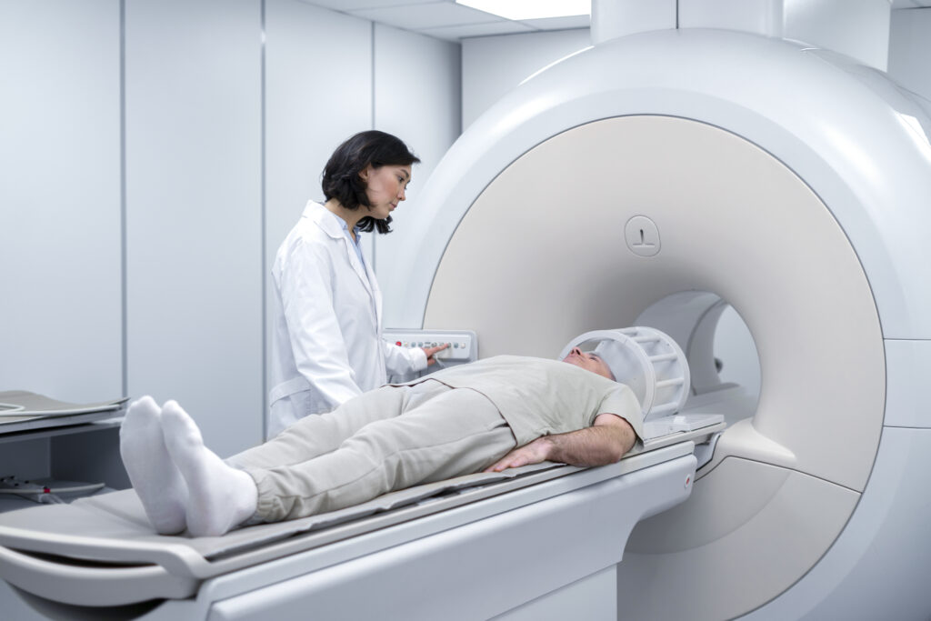
Magnetic Resonance Linear Accelerator, known as MR Linac, represents one of the most significant advancements in modern radiation therapy. This innovative system merges magnetic resonance imaging with a linear accelerator, enabling real-time visualization of tumors and nearby tissues during radiation delivery. The result is a powerful blend of precision, adaptability, and safety that is transforming how specialists approach cancer treatment within the field of medical technologies.
The Evolution of MR-Guided Radiotherapy
Conventional radiation systems relied heavily on static imaging performed before the treatment began. Once radiation delivery was underway, there was no mechanism to monitor subtle movements of the tumor or surrounding organs. This limitation often required specialists to apply wider safety margins, potentially affecting healthy tissue. The development of MR Linac addressed this challenge directly by allowing simultaneous imaging and radiation delivery.
By continuously capturing high-resolution magnetic resonance images during treatment, MR Linac provides clear visibility into how the tumor moves in real time. This is particularly valuable for cancers located in areas subject to motion such as the lungs, liver, pancreas, and abdomen. It allows specialists to adapt radiation beams dynamically, ensuring consistent targeting accuracy throughout every session.
MR Linac technology has therefore revolutionized the concept of image-guided radiotherapy, transforming it from a static process into a fully adaptive and responsive approach that personalizes therapy for each individual.
How MR Linac Works
MR Linac integrates two highly advanced systems: a magnetic resonance imaging unit and a linear accelerator. The MRI component delivers detailed soft-tissue images with unparalleled clarity, while the linear accelerator emits high-energy radiation beams precisely aimed at the tumor. These two systems function in harmony thanks to intricate engineering that prevents electromagnetic interference between them.
During each session, the MRI continuously scans the treatment area, allowing specialists to observe even the slightest motion of internal organs. If the tumor shifts due to breathing or digestion, the MR Linac immediately detects this change and adjusts the radiation beam’s position in real time. This capability ensures that the maximum dose is delivered to the tumor while minimizing exposure to healthy structures nearby.
The integration of imaging and radiation within one synchronized platform allows specialists to adapt each treatment session based on the current anatomy rather than relying solely on data acquired days or weeks earlier. This adaptability is the cornerstone of MR Linac’s precision.
Key Advantages of MR Linac Technology
The benefits of MR Linac extend far beyond simple imaging improvement. It represents a fundamental evolution in how radiotherapy is planned and delivered.
1. Real-Time Adaptive Therapy
MR Linac allows continuous monitoring of internal structures and dynamic adjustment of radiation beams. This ensures that each session is optimized based on the patient’s anatomy at that very moment, enhancing accuracy and consistency throughout the treatment course.
2. Enhanced Imaging Clarity
The MRI system integrated into MR Linac produces exceptional visualization of soft tissues, offering superior contrast compared to conventional imaging methods. This clarity helps specialists distinguish tumor margins more accurately, leading to improved target definition and better outcomes.
3. Protection of Healthy Tissue
Because MR Linac constantly tracks tumor motion, it can confine radiation precisely to the affected area, significantly reducing exposure to nearby healthy tissues. This results in fewer side effects and a smoother recovery experience for patients after therapy.
4. Personalization of Treatment
Each patient’s anatomy and tumor response are unique. MR Linac supports adaptive planning, enabling modifications to the radiation dose and target area during the treatment process. This individualized approach represents the next generation of personalized radiotherapy.
5. Efficient Workflow and Patient Comfort
The real-time imaging capability eliminates the need for additional scans between sessions, streamlining the workflow and reducing the overall treatment duration. Patients spend less time in treatment rooms while still benefiting from superior accuracy.
Applications Across Cancer Types
MR Linac technology is particularly beneficial for tumors located near organs that move during natural body processes. Cancers of the pancreas, liver, prostate, and lungs are among the most common cases where MR Linac’s precision makes a substantial difference.
For example, in abdominal tumors, small shifts caused by breathing can alter the target’s position by several millimeters. Conventional systems often compensate with larger safety margins, which may expose more healthy tissue to radiation. MR Linac, however, continuously adjusts the beam to match real-time motion, maintaining focus strictly on the tumor while sparing surrounding organs.
Additionally, MR Linac’s ability to visualize soft tissues without relying on contrast agents provides a safer, more comfortable experience for patients, especially those requiring multiple sessions.
Integration of Artificial Intelligence and Data Analytics
Recent developments are enhancing MR Linac’s capabilities even further through artificial intelligence and advanced data analytics. AI algorithms analyze imaging data to detect subtle changes in tumor shape, size, and response to therapy. These insights enable specialists to fine-tune treatment plans automatically, improving efficiency and outcomes.
Machine learning models can also predict how tumors will respond over time, helping clinicians decide when to adapt therapy schedules or modify doses. As AI continues to evolve, MR Linac systems are expected to become even more intelligent, integrating predictive analytics into daily clinical workflows.
Challenges and Future Perspectives
Despite its transformative benefits, MR Linac remains a complex and resource-intensive technology. Its installation requires specialized infrastructure, and the operation demands advanced training. However, ongoing developments are making the technology more accessible and efficient.
In the near future, it is expected that MR Linac systems will become more compact, user-friendly, and integrated with automated planning tools. Collaboration between engineering, imaging science, and oncology will continue to drive improvements, making MR-guided adaptive radiotherapy an increasingly standard practice worldwide.
As more centers adopt this technology, growing clinical evidence will further validate its advantages across different cancer types and patient populations.
The Broader Impact on Medical Technologies
MR Linac stands as a remarkable example of how innovation in medical technologies can reshape patient care. By combining imaging and therapy into a single adaptive process, it represents a shift toward precision medicine—an approach that tailors treatment to each individual’s unique biological and anatomical characteristics.
Beyond oncology, the engineering achievements behind MR Linac inspire further advances in multimodal imaging, robotics, and AI-driven treatment systems. These developments mark the beginning of a new era where imaging and therapy converge seamlessly, ensuring both safety and effectiveness.
Conclusion
MR Linac has redefined what is possible in radiation-based cancer therapy. Its ability to visualize, adapt, and deliver treatment simultaneously offers a level of precision that was previously unimaginable. By continuously monitoring anatomy in real time and personalizing therapy session by session, MR Linac provides an intelligent, patient-centered approach that sets new standards in medical technologies. As research and innovation continue, MR Linac will remain a cornerstone in the ongoing evolution of precise, adaptive, and efficient cancer treatment.

