

The human brain is the most complex organ in the body, serving as the central command center for all thought, emotion, and movement. It operates through a vast, intricate network of billions of nerve cells, or neurons, which communicate using tiny, precise electrical impulses. This constant electrical activity forms the foundation of our consciousness and bodily functions. Understanding the patterns of this activity is essential for assessing brain health and function.
In the realm of modern medical diagnostics, few technologies offer such a direct and non-invasive window into the functional status of the brain as the Electroencephalogram, or EEG. This sophisticated assessment tool is designed to listen to and record the “language” of the brain—its electrical rhythms.
Unlike imaging technologies that show the physical structure of the brain, an EEG provides a dynamic, real-time look at its function. This makes it an indispensable tool for evaluating a range of neurological conditions. It is a safe, painless procedure that captures the electrical “brainwaves,” providing invaluable information to specialists who are dedicated to understanding and supporting neurological wellness.
What is an Electroencephalogram (EEG)?
An Electroencephalogram is a functional assessment that measures, records, and interprets the electrical activity generated by the brain. When neurons communicate, they produce small electrical currents. When large groups of neurons synchronize and fire together, they create a detectable electrical rhythm known as a brainwave.
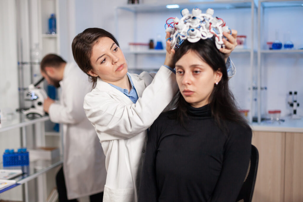
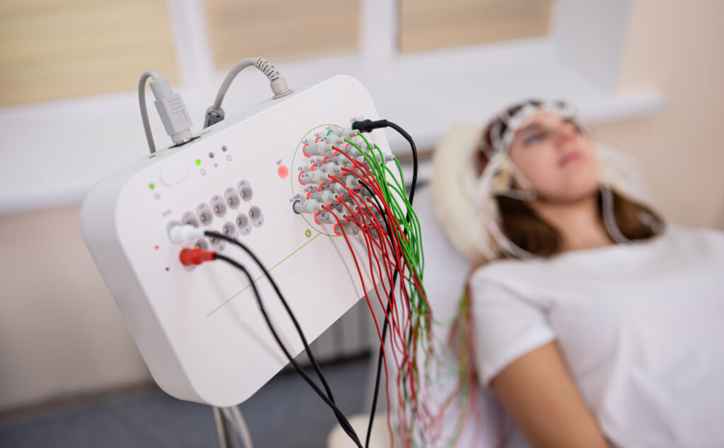
An EEG machine uses small, highly sensitive metal discs called electrodes. These sensors are carefully placed on the scalp to detect these brainwaves from different regions. The machine then amplifies these tiny signals and records them as a series of wavy lines on a computer screen.
This recording, the “encephalogram,” provides a visual representation of the brain’s activity over time. A specialist trained in neurophysiology can then analyze these patterns to identify signatures of normal brain function or detect abnormalities that may be associated with specific neurological conditions. It is a fundamental pillar of neuro-diagnostics, providing data that is often unattainable through other methods.
The Science of Brainwaves
The electrical patterns recorded by an EEG are not random; they are rhythmic, repetitive signals that change based on a person’s state of consciousness, alertness, or neurological status. These brainwaves are classified by their frequency (measured in cycles per second, or Hertz) and are associated with different states.
Types of Brainwave Patterns A specialist looks for several primary types of brainwaves during an EEG assessment:
- Beta Waves: These are fast-frequency waves. They are dominant when a person is awake, alert, mentally active, and focused on tasks or a conversation.
- Alpha Waves: These waves are slightly slower than beta waves. They are typically seen when a person is awake but in a relaxed, resting state, often with their eyes closed.
- Theta Waves: These are even slower waves, commonly associated with states of drowsiness, light sleep, or deep meditation.
- Delta Waves: These are the slowest and highest-amplitude brainwaves. They are the hallmark of deep, restorative sleep in adults.
The significance of an EEG lies not only in identifying these waves but in analyzing their context. For example, the presence of slow delta waves while a person is wide awake could indicate a disruption in brain function. Conversely, the absence of normal sleep patterns during a sleep assessment can be just as informative.
Why is an EEG Assessment Performed?
An EEG is an essential diagnostic tool used to gather information about brain function and to evaluate a variety of neurological symptoms. It is the primary assessment used to investigate sudden, unexplained changes in consciousness or behavior that may be related to brain activity.
Identifying Seizure Activity The most common and critical application of an EEG is in the evaluation of epilepsy and seizure disorders. A seizure is a sudden, uncontrolled electrical disturbance in the brain, often described as an “electrical storm.”
An EEG can capture the characteristic abnormal patterns associated with seizures, such as sharp “spikes” or “waves.” The information gathered helps specialists in several ways:
- It can help confirm that an event (like a blackout or staring spell) was, in fact, a seizure.
- It can help classify the type of seizure, which is critical for determining the most appropriate care path.
- It can help pinpoint the region of the brain where the seizure activity originates.
Evaluating Sleep Disorders The brain’s electrical activity changes dramatically and predictably as we move through the different stages of sleep. An EEG is a fundamental component of a polysomnogram, or sleep study. By monitoring brainwaves throughout the night, specialists can identify disruptions in the sleep-wake cycle, helping to evaluate conditions such as insomnia, parasomnias, and narcolepsy.
Monitoring Brain Function An EEG provides vital, real-time information about the overall health of the brain. It is often used to assess brain function in individuals who have experienced a significant head injury, a stroke, or a prolonged loss of consciousness (coma). It can also be used to evaluate encephalopathy, which is a general term for a change in brain function or structure caused by systemic or metabolic disturbances. In some cases, it may also provide supportive information in the evaluation of progressive neurodegenerative conditions.
The Patient Experience: What to Expect
The EEG procedure is a common, safe, and entirely painless assessment. It does not involve any needles, injections, or radiation. The electrodes only record activity; they do not transmit any electrical sensation to the individual.
Preparation for the Assessment Preparation for an EEG is straightforward and is designed to ensure the highest quality recording.
- Clean Hair: Individuals are typically asked to wash their hair the night before or the morning of the assessment. The hair should be completely dry and free of any products such as oils, sprays, gels, or conditioners, as these can interfere with the electrodes’ ability to detect signals.
- Medications and Diet: It is usually recommended to continue taking all prescribed medications unless specifically instructed otherwise. One should avoid consuming caffeine (from coffee, tea, or soft drinks) for several hours before the assessment, as it can affect the results.
- Sleep: For some EEG assessments, particularly those looking for seizure activity, the individual may be asked to be sleep-deprived. This might mean sleeping only for a few hours (or not at all) the night before. A tired brain is more likely to show abnormalities, making the assessment more informative.
During the EEG Procedure The assessment is performed by a specially trained EEG technologist.
- Measurement and Placement: The individual will sit or lie down comfortably. The technologist will first measure the person’s head and mark specific spots on the scalp with a soft crayon. This ensures the electrodes are placed precisely.
- Applying the Electrodes: The technologist will apply 20 or more small, metal-disc electrodes to the marked spots. These are held in place using a special adhesive paste or, in some cases, a stretchy elastic cap that has the electrodes embedded in it.
- The Recording: Once the electrodes are in place, the recording begins. The individual is simply asked to relax and lie still, first with their eyes open and then with their eyes closed. The technologist will monitor the recording from an adjacent room and may communicate via an intercom.
- Activation Procedures: To uncover potential abnormalities that are not present when the brain is at rest, the technologist may ask the individual to perform simple tasks. These are known as activation procedures and may include:
- Hyperventilation: Breathing deeply and rapidly for a few minutes.
- Photic Stimulation: Looking at a bright, flashing light (a strobe light) at different speeds.
- Drowsiness/Sleep: The person may be encouraged to fall asleep, as some brain abnormalities only appear during drowsiness or sleep.
After the Assessment A standard, or “routine,” EEG usually takes about 60 to 90 minutes, which includes setup time. The actual recording is often about 20 to 40 minutes. After the recording is finished, the technologist removes the electrodes. The conductive paste is harmless and is easily washed out of the hair with warm water.
Barring any specific instructions related to sleep deprivation, an individual can return to their normal daily activities immediately after the assessment.
Advanced EEG Modalities
While the routine EEG is the most common, medical technology has evolved to allow for more in-depth monitoring when needed.
- Ambulatory EEG: For individuals who experience events (like staring spells or seizures) infrequently, a standard 20-minute EEG may not capture one. An ambulatory EEG uses a small, portable recording device that the person wears for 24 to 72 hours while at home, allowing for long-term monitoring during normal daily life.
- Video-EEG Monitoring: Often considered the gold standard for evaluating complex seizure disorders, this assessment is performed over several days in a specialized monitoring suite. The individual is recorded on video 24/7 while the EEG is continuously running. This allows the care team to precisely correlate any physical events or symptoms seen on the video with the exact electrical activity happening in the brain at that same moment.
Commitment to Advanced Neuro-Diagnostics at Liv
At Liv, we are committed to providing access to the most advanced diagnostic technologies. The EEG is a cornerstone of our neurological assessment capabilities, offering profound insights into brain function.
We understand that technology is only one part of the equation. The true value of an EEG lies in the expertise of the team behind it. Our dedicated technologists are highly trained to conduct these assessments with precision and compassion, ensuring the comfort of every individual. Subsequently, our team of specialized neurologists, with extensive experience in neurophysiology, meticulously analyze and interpret the complex data. This collaborative, expert-driven approach ensures that every EEG assessment provides the clear and comprehensive information needed to guide the next steps in a person’s care journey.