

Ultrasonography is a widely used medical imaging technology that utilizes high-frequency sound waves to visualize internal structures of the body. By producing real-time images, this non-invasive modality allows specialists to assess organs, tissues, and blood flow with exceptional safety and efficiency. Ultrasonography is a cornerstone of modern medical technologies, valued for its diagnostic versatility and patient-centered approach.

The Evolution of Ultrasonography
Early imaging techniques, such as X-rays, provided structural information but were limited in assessing soft tissues or dynamic processes. Ultrasonography introduced a non-invasive, radiation-free method to evaluate organs and physiological functions in real time.
Advancements in transducer technology, image processing, and Doppler techniques have transformed ultrasonography into a powerful diagnostic tool. Modern systems offer high-resolution imaging, color flow visualization, and advanced 3D/4D capabilities, enhancing accuracy and clinical applications.
How Ultrasonography Works
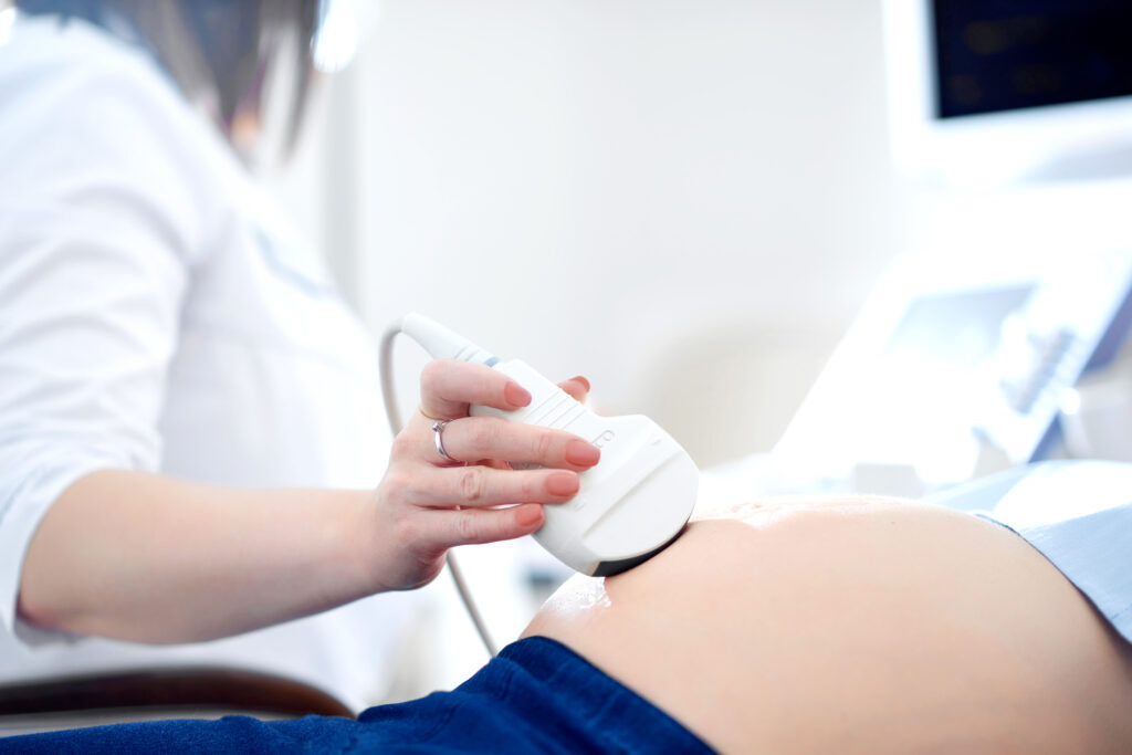
Ultrasonography employs a transducer that emits high-frequency sound waves into the body. These waves reflect off tissues and organs at varying rates depending on density and composition. The transducer then detects the returning echoes, which are processed by a computer to generate real-time images.
Different modes of ultrasonography provide specialized information:
- B-mode (Brightness mode): Produces two-dimensional grayscale images of anatomical structures.
- Doppler Ultrasound: Evaluates blood flow, velocity, and direction within vessels.
- 3D/4D Imaging: Offers volumetric and real-time dynamic imaging, often used in obstetrics and detailed organ assessments.
Key Advantages of Ultrasonography
- Real-Time Imaging
Ultrasonography provides immediate visualization, allowing dynamic assessment of organs, muscles, and blood flow. - Non-Invasive and Safe
The procedure does not use ionizing radiation, making it safe for repeated use and suitable for sensitive populations, including pregnant women and children. - Versatility Across Clinical Applications
It is applicable in obstetrics, cardiology, abdominal imaging, musculoskeletal assessments, and vascular evaluations. - Cost-Effective and Accessible
Ultrasonography is widely available, portable, and efficient, making it a practical tool in various healthcare settings. - Guidance for Interventions
It supports minimally invasive procedures, such as biopsies, fluid drainage, and catheter placements, by providing real-time visual guidance.
Applications Across Medical Fields
Obstetrics and Gynecology
Ultrasonography monitors fetal development, evaluates placental health, and assesses reproductive organs with high precision.
Cardiology
Echocardiography evaluates heart structure, function, and blood flow, aiding in diagnosis and management of cardiac conditions.
Abdominal Imaging
Liver, kidney, pancreas, and gallbladder assessments are efficiently performed using ultrasonography to detect lesions, stones, or fluid collections.
Vascular Studies
Doppler ultrasound assesses blood flow, identifies blockages, and evaluates aneurysms or varicose veins.
Musculoskeletal Imaging
Tendon, ligament, and muscle injuries can be visualized in real time, aiding diagnosis and treatment planning.
Integration of Advanced Imaging and AI
Modern ultrasonography systems incorporate advanced digital imaging, 3D/4D visualization, and AI-assisted analysis. Machine learning algorithms enhance image quality, detect abnormalities, and provide automated measurements, supporting faster and more accurate clinical decisions. AI integration also improves workflow efficiency and diagnostic confidence.
Challenges and Future Perspectives
While ultrasonography is highly versatile, image quality can be affected by patient anatomy, operator skill, and acoustic barriers. Proper training and experience are essential for optimal results.
Future developments include higher-resolution transducers, portable point-of-care devices, AI-driven diagnostics, and enhanced 3D/4D imaging capabilities. These innovations aim to expand the applications, accessibility, and accuracy of ultrasonography.
The Broader Impact on Medical Technologies
Ultrasonography exemplifies the combination of non-invasive imaging, real-time diagnostics, and advanced computational tools. By enhancing patient safety, improving diagnostic precision, and supporting a wide range of clinical applications, it plays a vital role in modern medical care and inspires further innovations in imaging technology.
Conclusion
Ultrasonography has revolutionized medical diagnostics by providing real-time, non-invasive, and highly versatile imaging. With ongoing technological advancements, including AI integration and 3D/4D imaging, ultrasonography continues to enhance diagnostic accuracy, patient safety, and clinical efficiency, solidifying its position as a cornerstone of modern medical technologies.